
A Look Into Imaging Technology for Neurological Disorders
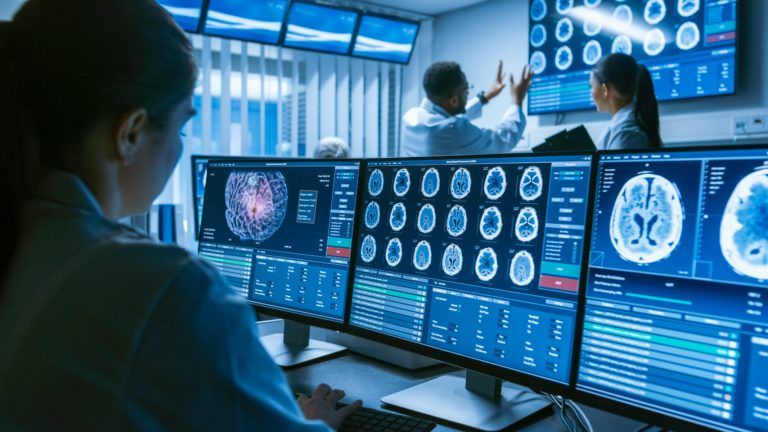
For hundreds of years the mysteries of the mind have been veiled behind a layer of hair, skin, and bone in the cradle of our selves that is our brain. Over 40 years ago this mystery was unveiled thanks to a tool being used by a chemist that was found to be able to make it possible to study the function and structure of the brain without requiring an autopsy. Today technologies like the MRI (Magnetic Resonance Imaging) are used to study the brain and learn more about neurological conditions.
The History Of The MRI
In the 1970’s a chemist by the name of Paul Lauterbur was enjoying his summer break from his teaching career when he a company that was at risk of bankruptcy reached out to him for help. The work the company was involved in was the sale of NMR’s, Nuclear Magnetic Resonance spectrometers that were used to figure out what chemical compounds are made of. It quickly came to his attention that these devices were capable of showing the difference between healthy and cancerous tissue in the body. It occurred to him that it may be able to produce pictures of organs and tissues found in a living patient.
The Structure and Function Of The Brain Revealed
Sure enough the NMR’s were in fact capable of revealing the structures he sought, and with some modifications the MRI was born and introduced to hospitals. The best part of the new innovation is it didn’t require the subject to be subjected to radiation such as computer tomography and x-rays, and yet were able to reveal the structure of the tissues and organs and what was happening inside them. Even more amazing is that the MRI could actually reveal activity happening in the brain.
Activity Within The Brain Isn’t Isolated
The brain is in constant motion, with multiple locations firing at any given time. Oxygen is used more aggressively by those areas of the brain that are active, resulting in more blood being sent to that area. The MRI is able to detect the difference between areas that are rich and poor in oxygen, and thus where activity was happening in the brain. As a result deeper research was able to be made into neurological conditions that affect the brain and reveal precisely what was going on in brains that were afflicted by them.
If you want to learn more about neurological imaging and work closely with a doctor who has been identifying these kinds of disorders for years, it’s time to call Dr. Diana Wilson at Neurosurgeon and Spine Fort Worth in Fort Worth, TX. With her team of diagnostic experts and specialists she’s been helping patients face the challenges of living with a neurological disorder and help them work their way to a life with higher quality of living. Don’t let life with a neurological disorder keep you from experiencing the fullest life possible, call today for a consultation to start working with the experts at Neurosurgeon and Spine Fort Worth.
Recent Comments